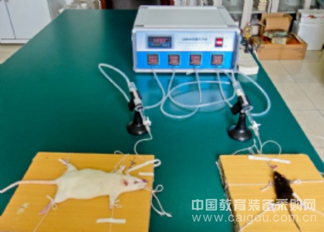
Ischemic retinal damage is a frequent and serious condition in ophthalmology, often resulting from inadequate blood supply to the retina. As a highly sensitive nerve tissue, the retina is particularly vulnerable to ischemia and hypoxia, especially when the central retinal artery—being a terminal vessel—is affected. This can lead to rapid and severe retinal injury, making it a critical issue in various eye diseases such as glaucoma and central retinal artery occlusion. Understanding the mechanisms behind retinal ischemia has long been a key focus in medical research, particularly within the field of ophthalmology. Investigating these mechanisms and evaluating the protective effects of potential drugs are essential for improving clinical outcomes and developing new treatment strategies. Creating an accurate and reliable experimental model for retinal ischemia is crucial for advancing research in this area. Currently, several common models are used, including elevated intraocular pressure, vascular ligation, and post-balloon injection of vasoconstrictors. Among these, the intraocular pressure elevation method is widely adopted due to its simplicity and effectiveness. It involves increasing the pressure inside the anterior chamber of the eye, which leads to retinal ischemia by compressing the vessels that supply the retina. In China, most researchers use self-made devices to create retinal ischemia models. These typically involve a sealed container filled with physiological saline and an inflatable system, where a rubber ball is manually squeezed to apply pressure. The pressure is monitored using a sphygmomanometer, but this process is labor-intensive, difficult to control precisely, and often results in inconsistent outcomes. The lack of an automated pressurizing device has been a major limitation in this field. To address these challenges, we developed a modular-designed animal retina pressure gauge. This device uses compressed air as the pressure source and incorporates a gas-liquid isolator to transmit the force. A pressure sensor detects the applied pressure, while a controller manages both pressure and duration. This automation ensures high precision (pressure range: 0–150 mmHg ± 2 mmHg), consistent results, and improved repeatability. It significantly reduces the workload for researchers, allowing them to focus on data analysis rather than manual operations. Additionally, the device is simple to construct, easy to operate, and cost-effective, making it a valuable tool for retinal ischemia research. Title: Animal Retina Pressure Gauge Completion Unit: Department of Pharmacology, School of Medicine, Shandong University Door Drapes,Bedroom Door Curtains,Macrame Door Curtain,Macrame Cotton Door Curtain Shandong Guyi Crafts Co.,Ltd , https://www.gyicraft.com
[Shandong University] Animal Retina Pressure Gauge