Stereo microscope operation method and application field Stereoscopic microscope, also known as "solid microscope" or "dissecting mirror", is a visual instrument with a positive stereoscopic effect and is widely used in various fields of biology, medicine, agriculture, forestry, industry and marine life. structure The magnification change of a stereo microscope is obtained by changing the distance between the intermediate mirror groups, and is therefore also referred to as a "Zoom-stereo microscope". With the requirements of the application, such as fluorescence, photography, camera, cold light source and so on. Characteristics 1. Using the most advanced Telescope (CMO) optical principle design to provide users with the sharpest image. 2. Perfect 3D image, providing clear, distortion-free images throughout the entire zoom range. 3. Wide field of view optical observation, Stemi 2000-C can provide you with a maximum field of view of 118mm. 4. With long working distance, Stemi 2000-C can provide you with working distance up to 286mm. Method of operation Stereomicroscopes use two independent optical pathways to generate three-dimensional optical images, so they are also called solid microscopes, dissecting microscopes, and low-power multiple optical microscopes. From the 1890s (1890) by American instrument engineer Horatio. Grino {the father (1805-1852) was invented by the famous American sculptor and writer Horatio Gino] and was first produced by Carl Zeiss in Germany, for scientific research, archaeological exploration, industrial quality Developments in areas such as control and biopharmaceuticals have had a positive impact. In order to maximize the efficacy of a stereo microscope, it is especially important to use a stereo microscope correctly. In order to let the users better operate the stereo microscope produced by Zhongwang Instrument, this article will share the operation flow of the stereo microscope with the article, specifically taking ZVM0745T, the best-selling product of Zhongwang Instrument, as an example. Steps step 1 Place the microscope on a platform that is comfortable for the operator, then turn on the reflected light (surface light), place a pattern on the microscope base, such as a coin, and rotate the microscope's zoom knob to the lowest multiple of 0.7X. The group finds the approximate focal plane (the best imaging plane) at 0.7X. Step 2 Adjust the eyelid of the eyepiece and adjust the diopter on the eyepiece to find the best focal plane at 0.7X. Step 3 Using the above method, gradually widen the multiple of the zoom knob, adjust the lifting group of the microscope appropriately, and gradually find the focal plane with a maximum magnification of 4.5X. During the adjustment process, use the more obvious reference points on the coin to compare the sharpness of the image. Different magnifications to observe the effect of coins Different magnifications to observe the effect of coins Step 4 Rotate the zoom knob to the lowest multiple of 0.7X. Maybe there will be some out of focus. In this case, please do not adjust the lift group to focus. Just adjust the diopter on the two eyepieces to adjust the eye (the diopter varies from person to person). ). At this point, the microscope is already in focus, that is, the microscope is changed from high magnification to low magnification, and the entire image is at the focal length. For the same sample, we don't need to adjust the other parts of the microscope. We only need to turn the zoom knob to easily zoom the sample. Folding and editing this paragraph application field The stereo microscope is easy to operate, the magnification is generally 7X-42X, and the maximum magnification is 180X. Stereoscopic microscopes are also the most widely used, and their main uses are as follows: 1. Research in zoology, botany, entomology, histology, mineralogy, archaeology, geology and dermatology. 2. In the textile industry, for the inspection of raw materials and cotton wool fabrics. 3. In the electronics industry, as a transistor spot welding, inspection and other operating tools. 4. Inspection of surface phenomena such as crack formation of various materials and corrosion of pore shape. 5. Equipment for machine tool tools, observation of working processes, inspection of precision parts, and assembly tools when manufacturing small precision parts. 6. Surface quality of lenses, prisms or other transparent materials, as well as quality inspection of precision scales. 7. Make a true and false judgment of the instrument money. 8. Widely used in textile products, chemical chemistry, plastic products, electronics manufacturing, machinery manufacturing, pharmaceutical manufacturing, food processing, printing, colleges and universities, archaeological research and many other fields. Troubleshooting Stereo microscopes have a wide range of applications in various fields of industry, agriculture and scientific research because of their many advantages. Stereomicroscope Stereomicroscope use. If some problems occur during use, you can solve them according to the actual situation. Common faults according to actual usage are: the field of view is blurred or there are dirt. Possible causes are dirt on the specimen, dirt on the surface of the eyepiece, dirt on the surface of the objective lens, and dirt on the surface of the work board. According to the actual situation, clean specimens, eyepieces, objective lenses and dirt on the surface of the work board can be solved. The reason why the double images do not overlap may be that the adjustment of the pupil distance may be correct, and the measures of correcting the pupil distance may be adopted. If the double images are not coincident, the visual adjustment may be incorrect, the visual adjustment may be performed again, or the magnification of the left and right eyepieces may be different. Check the eyepiece and reinstall the eyepiece of the same magnification. If the image is not clear, it may be that the surface of the objective lens is dirty, please clean the objective lens. If the image is not clear when zooming, it may be that the illuminance is not adjusted correctly and the focus is not correct. You can re-visit and adjust the focus. If the lamp burns frequently and the light flickers, it may be that the local line voltage is too high, the bulb burns out quickly and the wires are not connected properly. Please check the voltage and the microscope wire connection is firm, if not It may be that the bulb burned out quickly and the bulb can be replaced. The adjustment of the stereo microscope before use mainly includes: focusing, dioptric adjustment, interpupillary adjustment and lamp replacement. The following description will be respectively made. Focusing: Place the tabletop into the platen mounting holes on the base. When observing transparent specimens, use frosted glass platen; when observing opaque specimens, use black and white platen. Then loosen the fastening screw on the focus slide to adjust the height of the mirror body to a working distance that is approximately the same as the magnification of the selected objective lens. After adjustment, the fastening screws must be tightened. When focusing, it is recommended to use flat objects such as flat paper with printed characters, ruler, triangle, etc. Dioptric adjustment: first adjust the dioptric circle on the left and right eyepiece tubes to 0 position. Normally, look first from the right eyepiece tube. Turn the zoom handwheel to the lowest position, turn the focus handwheel and the diopter adjustment ring to adjust the specimen until the image of the specimen is clear, then turn the zoom handwheel to the highest position and continue to adjust until the image of the specimen is clear. So far, at this time, observe with the left eyepiece tube, if not clear, adjust the visual ring on the left eyepiece tube in the axial direction until the image of the specimen is clear. Pitch adjustment: Pull the two-eye lens barrel to change the exit distance of the two-eye lens barrel. When the user observes that the two circular fields of view in the field of view are completely coincident, it indicates that the pupil distance has been adjusted. It should be noted that due to the individual's vision and eye adjustment differences, different users or even the same user should use the same microscope at different times to adjust the focus to obtain the best observation. Whether replacing the light source bulb or replacing the light source bulb, be sure to turn off the power switch before disconnecting, and the power cord plug must be unplugged from the power outlet. When replacing the light source bulb, first unscrew the knurled screw of the upper light source box, remove the light box, then remove the bad bulb from the lamp holder, replace the good bulb, and then install the light box and knurled screw. When replacing the light source bulb, remove the frosted glass plate or black and white plate from the base, then remove the bad bulb from the lamp holder and replace it with a good bulb; then install the frosted glass plate or black and white plate. . When replacing the lamp, wipe the bulb with a clean soft cloth or cotton yarn to ensure the lighting effect. Imaging function The system of stereo microscope consists of an optical imaging system consisting of a metallographic microscope and a macro camera. Its purpose is to form an image of a metallographic sample or photograph. The stereo microscope can directly perform quantitative metallographic analysis on metallographic samples; the macroscopic camera is suitable for analyzing metallographic photographs, negative films and physical objects. In order to be able to store, process and analyze images with a computer, it is first necessary to digitize the image. A frame of image is composed of a distribution of different gradations, represented by mathematical symbols as j = j (x, y), x, y is the coordinates of the pixel on the image, and j is the gray value. Therefore, one frame of image can be represented by an m×n moment, each element of the moment corresponds to a pixel in the image, and the value of aij represents the gray value of the pixel belonging to the i-th row and the j-th column in the image. A CCD camera (Charge Coupled Device Camera) is an image digitizing device. The stereo microscope features on the metallographic sample are imaged on the CCD after the optical system and photoelectrically converted and scanned by the CCD, and then taken out as an image signal, amplified by an amplifier, quantized into gray scales, and then stored, thereby obtaining digital image. The computer sets the gray value threshold T according to the range of gray values ​​of the features to be measured in the digital image. For any pixel in the digital image, if its gray level is greater than or equal to T, white (gray value 255) is used instead of its original gray level; if less than T, black (gray value 0) is used instead of the original The gray scale, the stereo microscope can convert the gray image into a binary image with only black and white gray, and then perform necessary processing on the image, so that the computer can conveniently perform particle counting, area, and Peripheral measurement and other image analysis work. If pseudo color processing is used, 256 gray levels can be converted into corresponding colors, so that the details close to the gray level and its surrounding environment or other details can be easily recognized, thereby improving the image and facilitating the computer to process the multi-feature image. The company's main stainless steel water collector, tank bottom weld vacuum test box, reading instrument, eight-stage air microbial sampler, relay comprehensive tester, dual-wavelength scanner, coating thickness gauge, soil mill, tempered glass surface Flatness tester, sound sensor, portable electric water level gauge, network mouth flow meter, corrosion rate meter, portable scratch tester, freezing point tester, water quality tester, online ammonia tester, coating thickness gauge, coating Thickness gauge, soil pulverizer, digital thermometer, gas sampling pump, ceramic impact tester, automatic crystallization point tester, drug freezing point tester, reed switch tester, constant temperature water bath, gasoline root turn, gas Sampling pump, tempered glass tester, water quality tester, PM2.5 tester, inhalable particulate matter detector, high frequency heat sealing machine, strain control triaxial instrument, milk somatic cell detector, helium gas concentration detector, soil moisture conductance Rate tester, field strength meter, collection box, color ratio tester, capillary water absorption time measuring instrument, redox potentiometer vibrometer, carbon monoxide carbon dioxide detection , CO2 analyzer, oscillopolarograph, slime content tester, car starter power supply, automatic potentiometric titrator, portable thermometer, zirconia analyzer, reed switch tester, precision conductivity meter, TOC water quality analysis Instrument, microcomputer plasticity tester, wind direction station, automatic spotting instrument, soil oxidation reduction potentiometer, digital thermometer, portable total phosphorus tester, corrosion rate meter, constant temperature water bath, residual chlorine detector, free expansion rate meter , centrifuge cup, concrete saturated vapor pressure device, particle strength tester, Gauss meter, automatic film coating machine, safety valve grinding tool, weather station, kinesthetic orientation instrument, dark adaptation instrument, smell collector, rain gauge, four in one Gas analyzer, emulsion concentration meter, dissolved oxygen meter, temperature measuring instrument, thin layer planking device, temperature recorder, aging meter, noise detector, constant temperature and humidity chamber, split resistivity tester, initial viscosity and holding viscosity Tester, infrared carbon dioxide analyzer, hydrogen lamp, kinesthetic orientation instrument, constant temperature animal operating table, cooling fan, grease acid value detector, viscosity meter, colony counter, Weather station, rain gauge, Kjeldahl nitrogen analyzer, fluorescent whitening agent, flat grinding instrument, fuel cell characteristic comprehensive tester, dissolving instrument, online PH meter, on-site dynamic balance meter, high vacuum valve, ultrasonic liquid level transmitter , Abbe refractometer, water quality rapid detector, sludge ratio group measuring device, scanner, nitrogen blowing instrument, personal dose alarm device, the company adhering to the "customer first, forge ahead" business philosophy, adhere to the "customer first" The principle is to provide quality services to our customers. Welcome patrons!
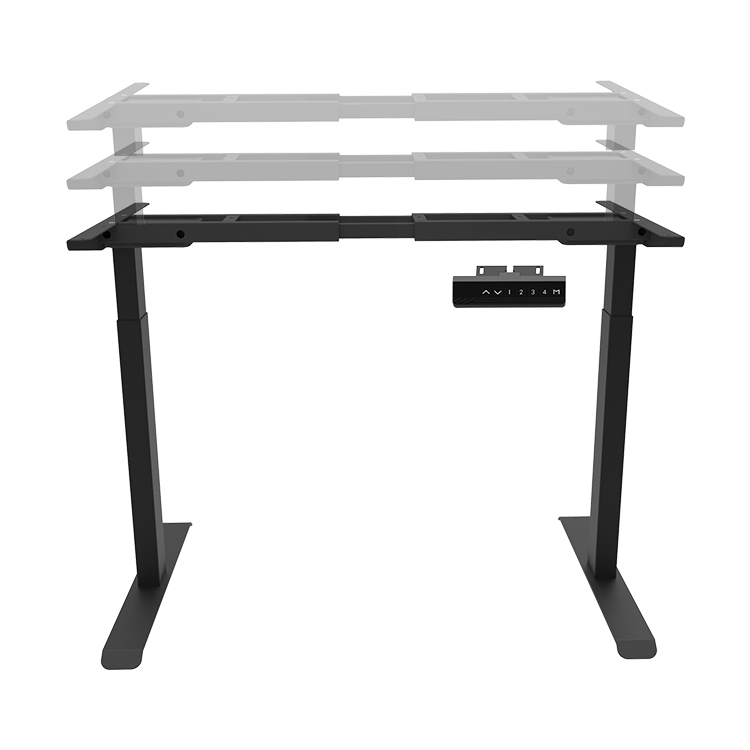
1. Sit Stand Workstation Lift freely, can stand can sit alternate office, stand sit alternate do not hinder busy business at hand.
2. It's easy to operate, just press the button to achieve the purpose of alternate standing and sitting.
3. Memory function storage: that is, the user can adjust the function key to a suitable height according to his own height, and adjust the function key to a fixed position to facilitate the fixed position and use.
4. The appearance is simple and generous, which is in line with the international office trend and different from the traditional desk style which is dull and not easy to move.
5. It is beneficial to the physical and mental health of office workers, increases work efficiency, enlivens the workplace atmosphere and makes the workplace atmosphere more active.
Sit Stand Workstation,Stand up desk workstation,Sit Stand Desk Workstation,Adjustable Work Tables,Sit Stand Laptop Desk Suzhou Uplift Intelligent Technology Co., Ltd , https://www.uplifting-desk.com
Stereo microscope operation method and application field